drnbansal@ymail.com
Phone : +918447542860
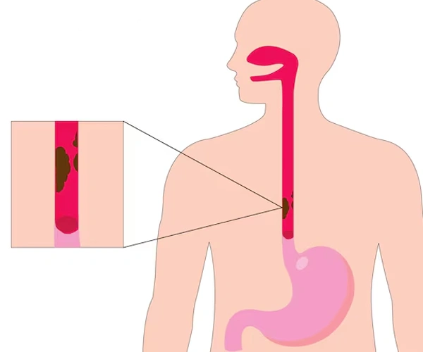
If you are searching for the best Granular Cell Tumor of Esophagus Treatment in Janakpuri, accurate diagnosis and expert gastroenterology care are important for managing rare tumors of the food pipe. Granular Cell Tumor of Esophagus is an uncommon tumor that develops from specialized nerve-related cells within the esophageal wall. Most granular cell tumors are benign (non-cancerous), but careful evaluation is required because some cases may show aggressive features.
These tumors are often discovered incidentally during upper GI endoscopy performed for other digestive symptoms. Larger lesions may cause swallowing difficulty, chest discomfort, or a sensation of food getting stuck.
At Dr. Naresh Bansal's Gastro & Liver Clinic, patients receive advanced diagnostic evaluation, including endoscopy and endoscopic ultrasound (EUS), along with personalized treatment planning for esophageal tumors and other gastrointestinal conditions.
What is Granular Cell Tumor of Esophagus?
Granular Cell Tumor of Esophagus is a rare type of tumor that arises from Schwann cells, which are cells associated with nerves.
These tumors usually develop in the inner layers of the esophageal wall and are generally:
- Slow-growing
- Small in size
- Benign in most cases
- Detected accidentally during endoscopy
Although most esophageal granular cell tumors are harmless, evaluation is important to differentiate them from other esophageal growths and determine appropriate management.
Causes and Risk Factors of Granular Cell Tumor of Esophagus
The exact cause of granular cell tumors is not clearly understood.
Possible factors involved include:
- Abnormal growth of Schwann cells
- Genetic changes affecting cell development
- Local tissue changes
These tumors are not usually linked to:
- Diet
- Acid reflux
- Smoking
- Infections
Granular cell tumors can occur in adults of different age groups and may affect both men and women.
Symptoms of Granular Cell Tumor of Esophagus
Many small granular cell tumors do not produce symptoms and are found during routine investigations.
When symptoms occur, they may include:
- Difficulty swallowing (dysphagia)
- Feeling of food getting stuck
- Chest discomfort
- Pain while swallowing
- Regurgitation
- Heartburn-like symptoms
- Unexplained weight loss in rare cases
Symptoms usually depend on:
- Size of the tumor
- Location in the esophagus
- Effect on food passage
Complications of Granular Cell Tumor of Esophagus
Although most tumors are benign, possible concerns include:
Swallowing Difficulty
Larger tumors may interfere with normal food movement.
Growth of the Lesion
Some tumors may increase in size and require intervention.
Diagnostic Uncertainty
Some submucosal tumors require detailed evaluation to confirm their nature.
Rare Malignant Transformation
A very small percentage of granular cell tumors may show malignant features, making proper assessment important.
How is Granular Cell Tumor of Esophagus Diagnosed?
Diagnosis requires detailed evaluation of the tumor structure and tissue characteristics.
Upper GI Endoscopy
Endoscopy is usually the first test used to identify the lesion.
It helps:
- Locate the tumor
- Assess size and appearance
- Evaluate the esophageal lining
Endoscopic Ultrasound (EUS)
EUS provides detailed images of the layers of the esophageal wall.
It helps determine:
- Depth of tumor involvement
- Layer of origin
- Tumor size
- Internal characteristics
Biopsy and Histopathology
Tissue examination may be required to confirm diagnosis.
A biopsy helps identify:
- Granular cell features
- Benign or suspicious characteristics
- Need for further treatment
Imaging Tests
CT scan or other imaging may be recommended in selected cases to assess larger lesions.
How is Granular Cell Tumor of Esophagus Treated?
Treatment depends on:
- Tumor size
- Symptoms
- Growth pattern
- Histopathology findings
Observation and Monitoring
Small, asymptomatic tumors with benign features may be monitored through regular follow-up.
Endoscopic Removal
For suitable small lesions, endoscopic removal may be considered.
Benefits include:
- Minimally invasive approach
- No external incision
- Faster recovery
Surgical Removal
Surgery may be recommended when:
- The tumor is large
- Symptoms are significant
- The lesion has suspicious features
- Complete removal is required
Treatment Procedure
Step 1 – Initial Consultation
A detailed assessment of symptoms, medical history, and previous investigation reports.
Step 2 – Diagnostic Evaluation
Endoscopy, EUS, and biopsy may be performed to evaluate the tumor.
Step 3 – Tumor Assessment
The size, location, and characteristics of the lesion are analyzed.
Step 4 – Treatment Planning
A personalized approach is selected based on diagnosis and patient condition.
Step 5 – Follow-Up Monitoring
Regular follow-up helps ensure proper management and detect any changes early.
Benefits of Early Granular Cell Tumor Treatment
Seeking timely Granular Cell Tumor of Esophagus Treatment in Janakpuri provides several benefits:
- Accurate diagnosis
- Identification of tumor characteristics
- Prevention of complications
- Relief from swallowing symptoms
- Early detection of suspicious changes
- Appropriate treatment planning
- Improved digestive health management
- Better long-term outcomes
Why Choose Dr. Naresh Bansal?
Dr. Naresh Bansal is an experienced Gastroenterologist specializing in gastrointestinal disorders, esophageal conditions, and advanced endoscopic procedures.
Patients choose Dr. Naresh Bansal because of:
- Extensive experience in gastroenterology
- Expertise in evaluating esophageal tumors
- Advanced endoscopic diagnosis
- Experience with EUS-based evaluation
- Accurate assessment of submucosal lesions
- Personalized treatment planning
- Evidence-based medical approach
- Focus on minimally invasive procedures
- Comprehensive digestive healthcare
- Long-term follow-up support
Frequently Asked Questions (FAQs)
1. What is a granular cell tumor of the esophagus?
A granular cell tumor of the esophagus is a rare tumor that develops from nerve-related cells in the esophageal wall. Most cases are benign.
2. Is granular cell tumor of the esophagus cancerous?
Most granular cell tumors are non-cancerous, but proper evaluation is required to rule out aggressive features.
3. What symptoms can an esophageal granular cell tumor cause?
Symptoms may include difficulty swallowing, chest discomfort, and the sensation of food getting stuck.
4. How is granular cell tumor diagnosed?
Diagnosis usually involves endoscopy, EUS, and biopsy or tissue examination.
5. Does every granular cell tumor require removal?
No. Small benign tumors without symptoms may only require monitoring, while larger or suspicious lesions may need removal.

Dr Naresh Bansal.All Rights Reserved © 2026